Calcium pyrophosphate dihydrate (CPPD) crystals but not basic calcium phosphate (BCP) crystals induce syndecan-4 expression in cartilage
S. Nasi (1), J. Bertrand (2), M. Bollman (2), R. Stange (1(), T. Pap (1)
Affiliation(s):
1. University Hospital Münster, Germany,
2. University Otto Von Guericke Magdeburg, Germany
Status: Chondrocalcinosis is a painful rheumatic condition caused by the deposition of calcium pyrophosphate dihydrate crystals (CPPD) in joint tissues, and especially in cartilage. It is known that CPPD crystals cause inflammation and degenerative changes in joint, but the underlying mechanisms remain poorly understood. In particular, nothing is known about how these crystals regulates transmembrane heparan sulphate proteoglycans (HSPGs). Our attention focused on one family of HSPGs called syndecans as they have important roles both as adhesion molecules, by mediating chondrocyte-extracellular matrix interactions, and as modulators of intracellular signaling triggered by cytokines and growth factors.
Objectives: The aim of this study was to evaluate how CPPD crystals modulate syndecan expression in chondrocytes and in cartilage, and how this modulation can be ultimately linked to cartilage damage during chondrocylcinosis.
Methodology: Murine chondrocitic ATDC5 cells were stimulated with 0,1 ng/ml CPPD crystals or with 0,1 ng/ml basic-calcium phosphate crystals (BCP), a family of calcium-containing crystals found in other rheumatic conditions such as osteoarthritis (OA). Cytotoxicity was evaluated by lactate dehydrogenase (LDH) release in the supernatant at 30 minutes, and 3, 6, 24 hours after stimulation. At the same time-points, mRNA expression levels of syndecans (Synd-1, -2, -3, -4) and of matrix-degrading enzymes (Mmp-3, -9, -13; Adamts-4, -5) was analysed by qRT-PCR. Finally, Syndecan-4 protein expression was studied by immunohistochemistry (IHC) in cartilage samples of patients with chondrocalcinosis and in samples of patients with severe OA without chondrocalcinosis as control.
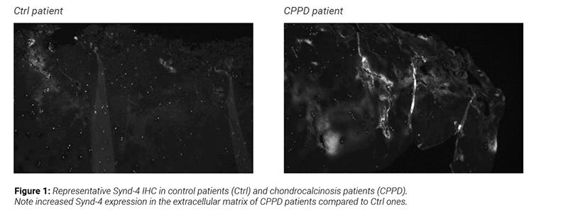
Findings: LDH assay revealed no increased cytotoxicity by CPPD or BCP at any time-point. qRT-PCR indicated that CPPD crystals but not BCP crystals induced Synd-2 and -3 upregulation at 30 minutes after stimulation and Synd-4 upregulation at 3 hours, while no modulation of syndecans was seen at later time-points. CPPD also induced Adamts-4 expression at 3 hours after stimulation, and Mmp-9 expression at 3 and 6 hours. The expression of the other matrix-degrading enzymes was not affected. Human chondrocalcinosis cartilage exhibited enhanced Synd-4 expression compared to severe OA cartilage containing BCP calcification. Interestingly, Synd-4 expression was observed in the extracellular matrix but not on cell membrane, suggesting that maybe Synd-4 undergoes shedding (Fig. 1).
Conclusions: BCP and CPPD crystals seem to trigger differential effects in terms of modulation of syndecans in chondrocitic cells. CPPD crystals induce Synd-4, Adamts-4 and Mmp-9 which are not induced by BCP crystals. It remains to be clarified whether the two events are interlinked. In particular, further studies are required to investigate if Adamts-4 and Mmp-9 are involved in Synd-4 shedding or if vice versa Synd-4 regulates Adamts-4 and Mmp-9 activation and downstream cartilage breakdown in chondrocalcinosis.
